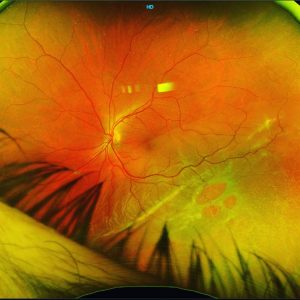
Here’s a great teaching photo of an unique rhegmatogenous retinal detachment.
On first inspection, it looks like a round hole “retinogenic” retinal detachment however there’s clues to show this is not the case.
1) There’s peripheral cystoid degeneration peripherally
2) There’s a mix of small round holes and larger rolled edge holes
3) The largest hole actually has a superficial/inner retinal vessel running over it!
This is a retinoschisis retinal detachment (RSRD) with inner and outer leaf retinal breaks leading to a full thickness retinal break and subsequent retinal detachment.
#vitreoretinal #retina#ophthalmology#vitreoretinalsurgery #eyedisease #retinasurgery #retinalsurgery #eyesurgery #ophthalmologist #eyedoctor #eyedisease #eyehealth #optometry #optometrist #optometrystudent #yamane #canabrava #vitrectomy #retinaldetachment #macula #laser #yamane #macula #medstudent #medicalstudent #goldcoasteyes #outlookeye #goldcoasteyesurgeon #goldcoastoptom