Our first “Case of the Week”!!
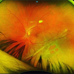
These are colour photos and fluoroscein angiograms of a 30yo female who presents with progressive acuity loss OS.
On examination she has Angioid Streaks (AS) radiating from the disc and is now involving her macula OS, hence the deteriorating vision.
She also has Peau d’Orange appearance of the fundus (yellow stars) which is pathognomonic of Pseudoxanthoma Elasticum (PXE).
Angioid Streaks are bilateral, narrow irregular “Jagged” or “vessel-like” lines deep to retina that radiate from the ONH. They form from full thickness breaks in a weakened and calcified Bruch’s with disruption of overlying RPE.
Tend to develop in patients with certain systemic disorders marked by extensive calcification of Bruch’s elastic layer.
Complications/Visual Loss occur in over 70% of patients through either
a. CNV – Most common cause of visual loss
b. Choroidal Rupture – May occur following trivial trauma eg rubbing eye
c. Foveal involvement of streak
Our favourite mnemonic for causes of AS was PEPSI MAX -
Pseudoxanthoma Elasticum
Ehler-Danlos Syndrome
Paget’s Disease of bone
Sickle Cell Disease and other haemoglobinopathies
Idiopathic (~50% of angioid streaks)
Myopia
Acromegaly, Abetalipoproteinaemia
toXic – Lead poisoning
#retina #ophthalmology #vitreoretina #ophthalmologist #eyedoctor #optometry #optometrist #optometrystudent