Case Studies
Guess the diagnosis
Not often they present like the textbooks! Had to share.Guess the diagnosis and what’s your next steps?!
OCTs and OCTAs of a Neovascular (wet) Age-Related Macular Degeneration (nARMD)
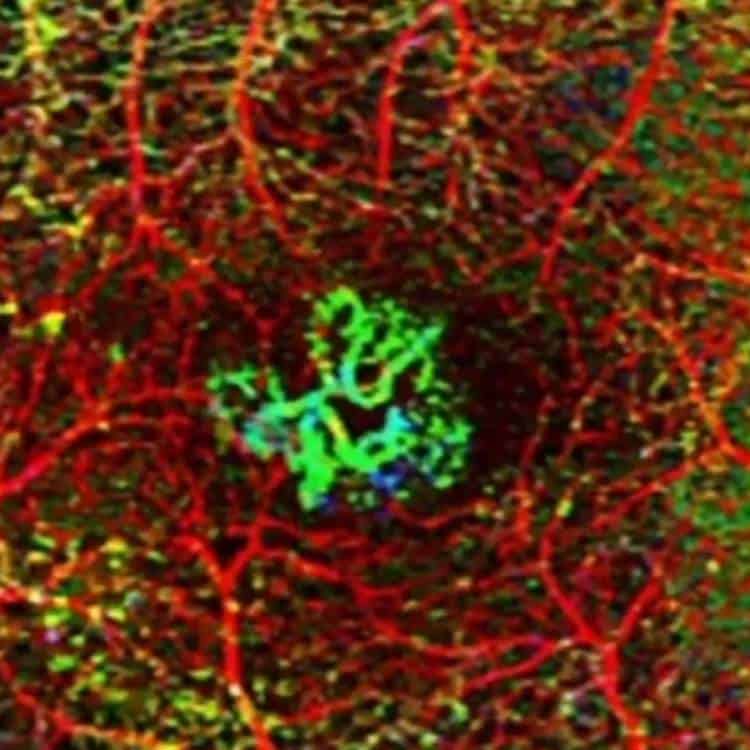
Here’s a series of OCTs and OCTAs of a neovascular (wet) Age-Related Macular Degeneration (nARMD) patient who underwent treatment with intravitreal anti-VEGF injections of Aflibercept. The initial (2nd image) OCT cross-sectional (B-scan) demonstrates subretinal and intraretinal fluid (SRF & IRF) with subfoveal haemorrhage. The third image is the OCT B-scan with flow overlay showing […]
Rhegmatogenous Retinal Detachment
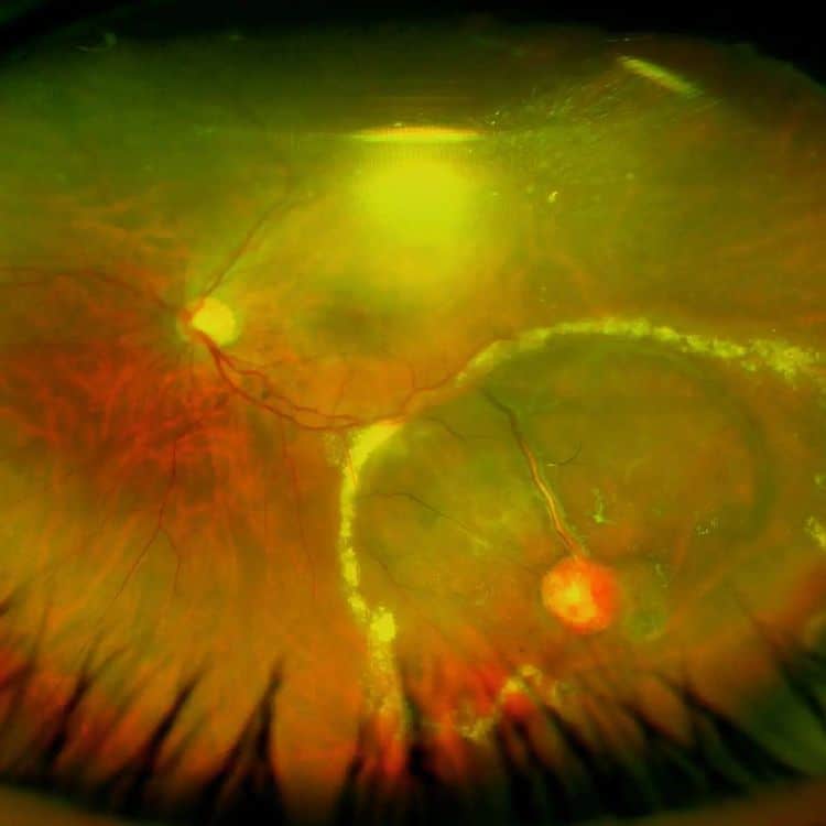
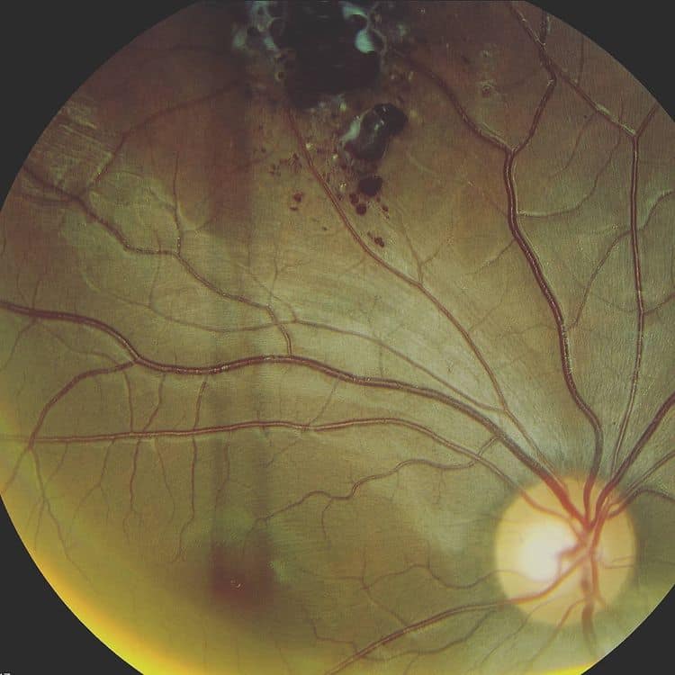
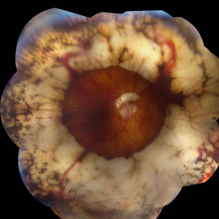
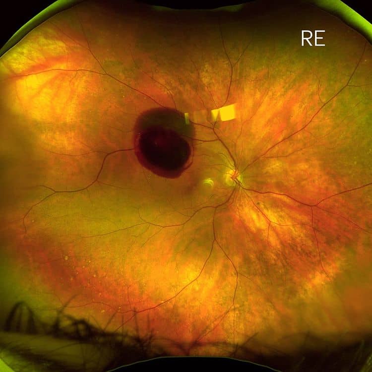
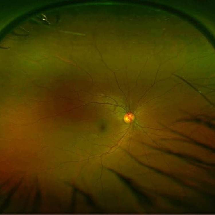
Here’s a great teaching photo of an unique rhegmatogenous retinal detachment. On first inspection, it looks like a round hole “retinogenic” retinal detachment however there’s clues to show this is not the case. 1) There’s peripheral cystoid degeneration peripherally2) There’s a mix of small round holes and larger rolled edge holes3) The largest hole […]
PDR TRD Repair
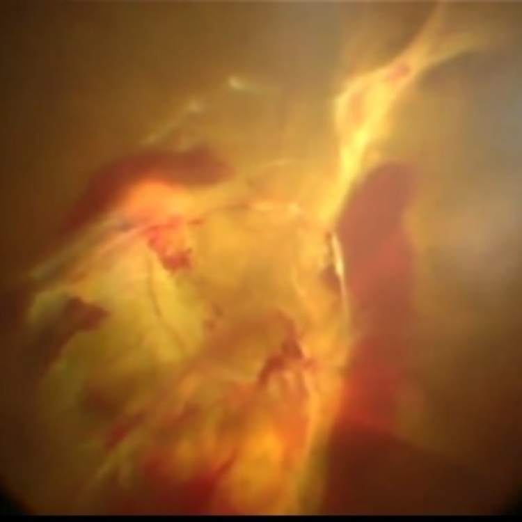
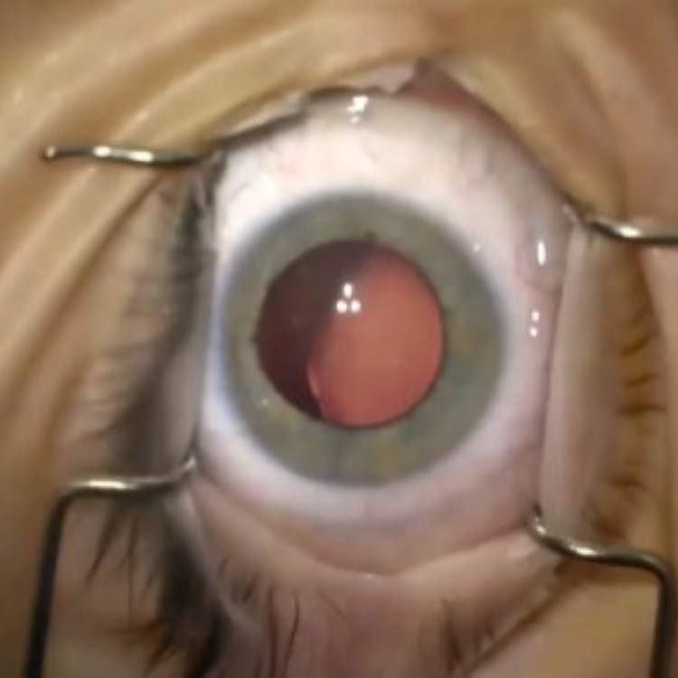
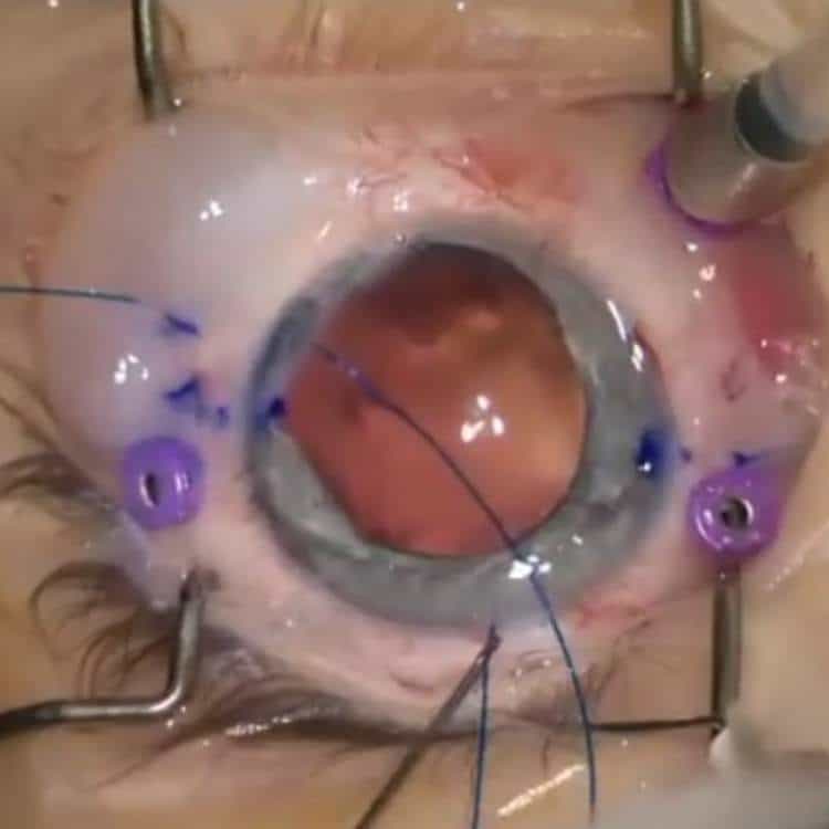
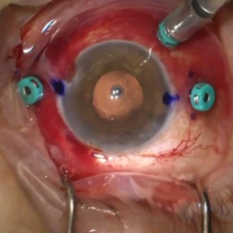
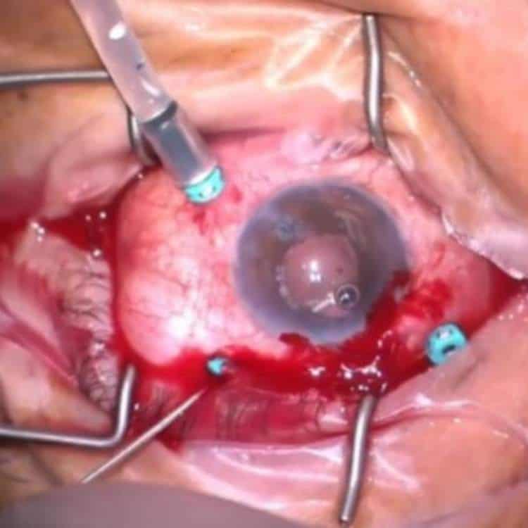
This is a short video of the eye in the preceding diabetic post. The video demonstrates the intricate and delicate work necessary to achieve successful reattachment of the retina. Once attached, adequate panretinal photocoagulation (PRP) laser can be performed to reduce the VEGF produced from ischaemic retina that drives the proliferative neovascularisation.
Proliferative Diabetic Retinopathy Tractional Retinal Detachment (PDR TRD)
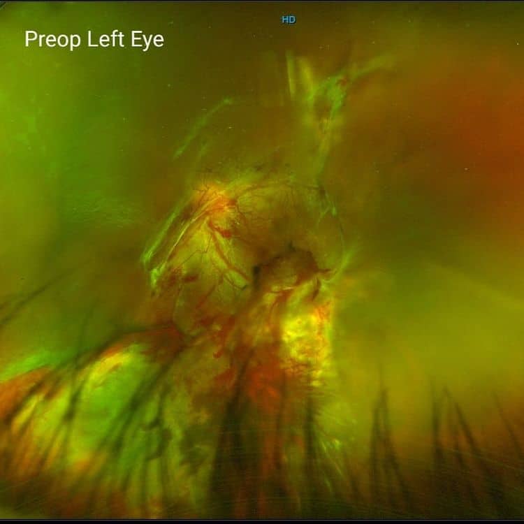
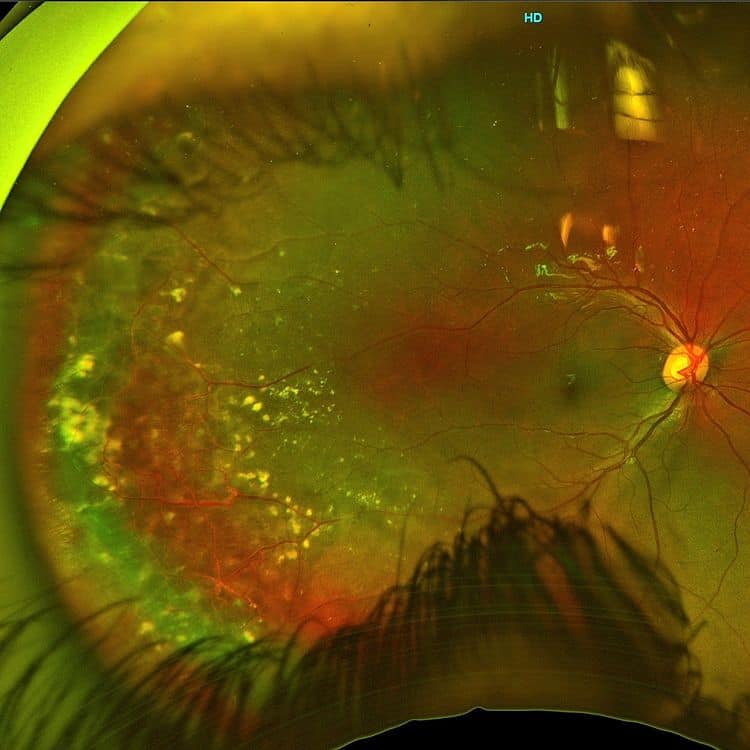
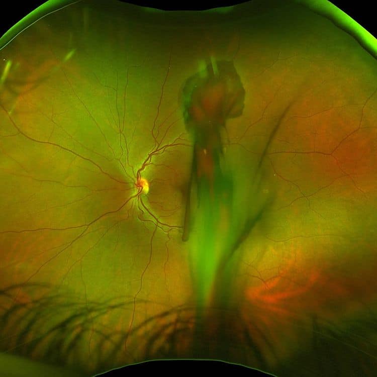
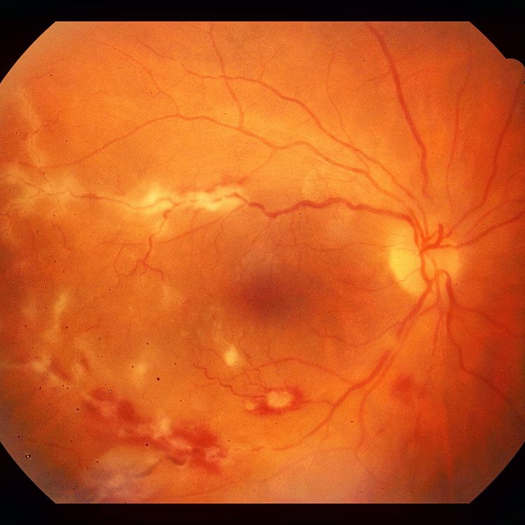
Another week, another proliferative diabetic retinopathy tractional retinal detachment (PDR TRD). We had 2 nasty ones this week but here’s a recent patient with an updated postoperative photo at 6months post-op. Preoperative photo shows a large posterior tractional detachment with overlying vitreous haemorrhage in their left eye. This patient actually presented due to sudden loss of […]
2021 Optometric Education Series
Outlook Eye Specialists are back for the second instalment of our 2021 Optometric Education series. Please come along to the second instalment of Outlook’s education series at our SOUTHPORT location. This year, we have split presentations to allow for a deeper dive in to topics, more Q&A and keeping sessions within a manageable timeframe. The […]
Retinal Capillary Haemangiomas – Peripheral and Juxtapapillary
Here’s a series of photos from separate patients demonstrating the 2 types of Retinal Capillary Haemangiomas – Peripheral and Juxtapapillary. Retinal Capillary Haemangiomas are vascular hamartomas that can not only cause slowly progressive visual problems but may also be associated with Von-Hippel Lindau (VHL) disease. RCHs can be isolated, multiple or bilateral. Peripheral RCHs appear […]
Flanged Intrascleral Fixation of Single-piece IOL
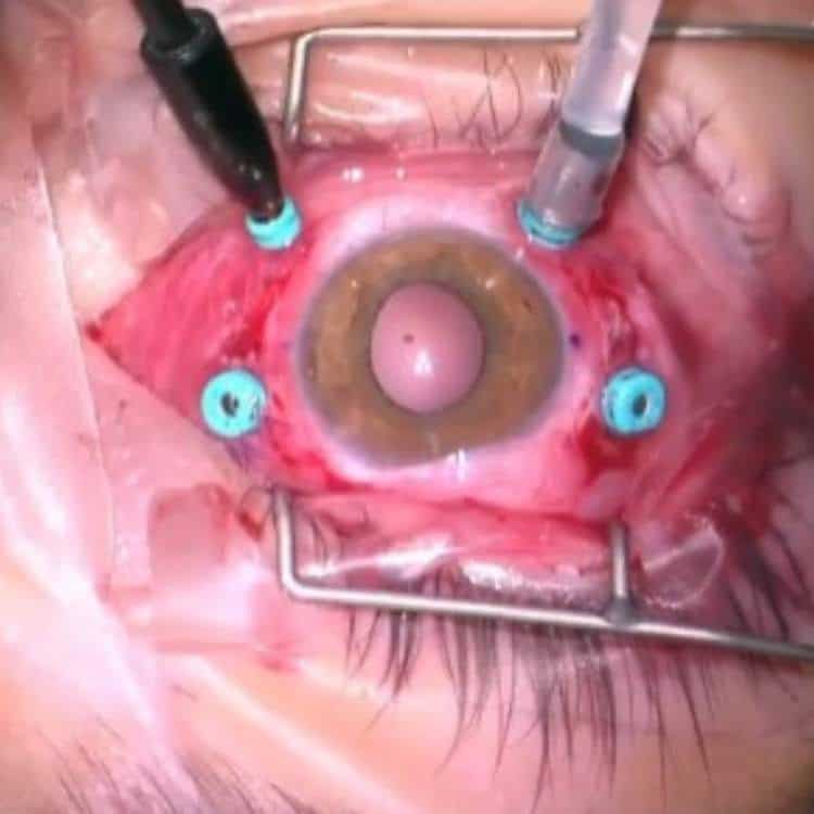
Flanged intrascleral fixation of a toric extended vision Alcon Vivity IOL in a paediatric patient with ectopic lentis from Marfans. We used modifications to the 4 flange technique originally described by @dr_canabrava. The plan was for in-the-bag placement with sutured capsular tensions segments and rings however there was over 270 degrees of zonular dehiscence and scleral […]
Coats’ Disease
This was a 7 year old male that was referred for treatment of their increasing peripheral exudation despite previous laser treatment. This patient had typical findings of early Coats’ Disease (Stage 2A) with mostly temporal and peripherally located vessel thickening/dilatation, telangiectasias, bulb-like aneurysms as well as capillary drop out clearly seen on angiography. This patient […]
Idiopathic Retinal Cavernous Haemiangiomas
“Raisin” awareness of this rare finding..Here’s a young male that was referred for possible BRVO however on examination it’s clear that this wasn’t the case!This was an incidental finding in a 25yo male with no ocular, medical or family history. After thorough systemic work-up and investigation including neuroimaging, he was diagnosed with idiopathic Retinal Cavernous […]
Metallic Intraocular Foreign Body (IOFB)
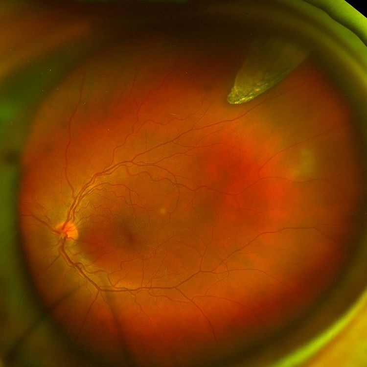
Close Call!! Here’s a patient with an metallic intraocular foreign body (IOFB) which occurred from hammering metal at work. This patient had the IOFB enter through the sclera and managed to embed itself in the vitreous cavity with no damage to the cornea, lens, macular or optic nerve.Fortunately his visual axis was spared from […]
X-Linked Retinoschisis (XLRS)
Here’s a case of X-Linked Retinoschisis (XLRS)This is an inherited disorder affecting males, generally diagnosed in childhood due to an inherited mutation in RS1 gene (which encodes for a protein involved in intercellular adhesion and organisation – Retinoschisin). It leads to bilateral reduced central vision which may result in nystagmus or strabismus. Vision generally stabilises […]
Retinal Aterial Macroaneurysm
Fun photo.. Here’s a case of a Retinal Aterial Macroaneurysm (RAMA) that ruptured following blunt trauma leading to a gravitational vitreous bleed. This patient went on to have a dense vitreous haemorrhage requiring vitrectomy. Vision was completely restored following surgery. #retina #ophthalmology #vitreoretinalsurgery #eyedisease #retinasurgery #eyesurgery #ophthalmologist #eyedoctor #optometry #optometrist #optometrystudent #vitrectomy #retinaldetachment #macula #laser […]
Optic Disc Pits
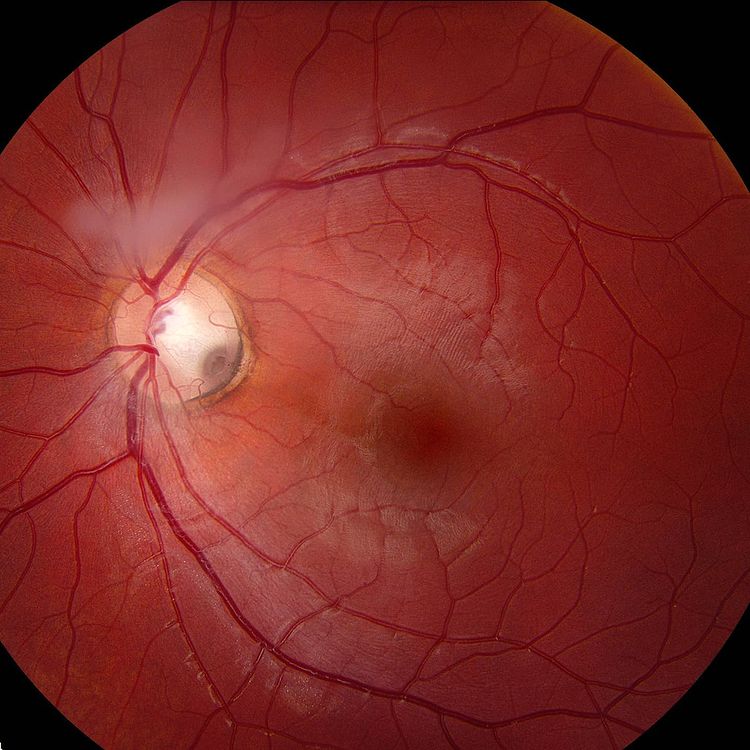
These rare congenital defects can be found in all ages and both sexes. Their origins are unclear however they are most likely due to closure failure of the optic fissure.They are usually unilateral ~90% of the time and solitary however multiple pits can be observed in the same nerve. Patients may be asymptomatic or have […]
Serpiginous Choroidopathy
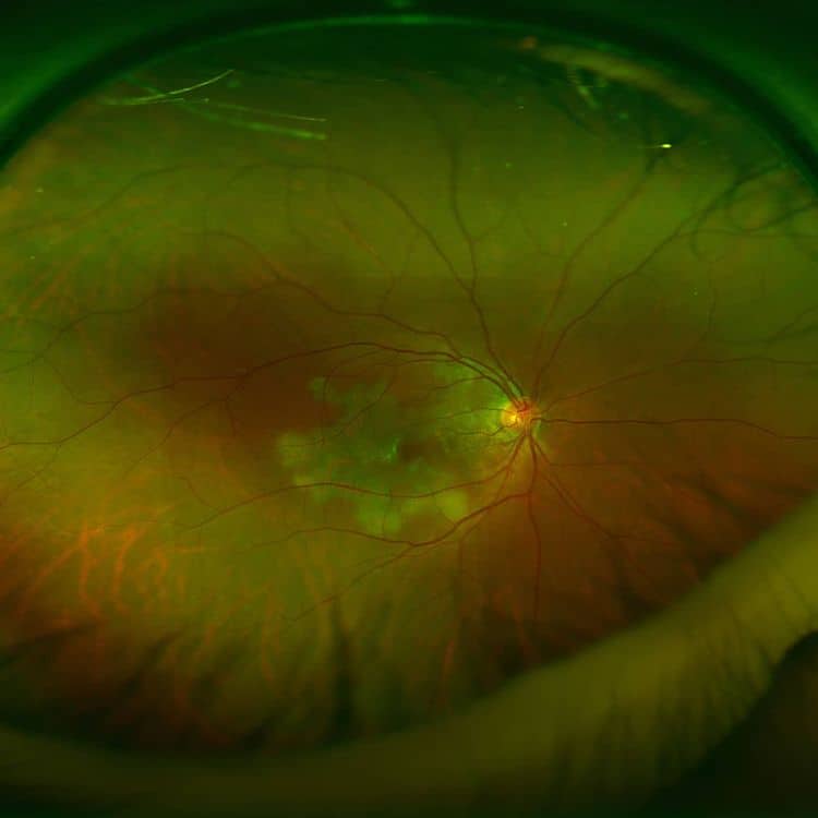
This young 27yo female presents with sudden loss of vision in the right eye. Vision dropped to 6/12 (20/40). Anterior examination was unremarkable. Posterior examination revealed large areas of retinal whitening emanating from the optic disc of the right eye involving the macula. This patient has Serpiginous Choroidopathy, which is part of the Placoid […]
Outlook Optometry Event 2020
Outlook Optometry Event 2020 with @theyotclub @outlookeye were finally able to have our first academic and educational event for 2020. It’s been a bit of crazy year so we decided to make it an extra special event on board the @theyotclub. Food, drinks and a few sunset talks on the Gold Coast Broadwater, sounds like a pretty amazing […]
Gyrate Atrophy
Here’s an advanced case of Gyrate Atrophy (Orthinine Aminotransferase Deficiency).This 67yo woman has suprisingly stable symptoms of poor night vision (nyctalopia) and reduced peripheral visual field despite the diagnosis of Gyrate Atrophy. Gyrate atrophy is an autosomal recessive condition caused by a deficiency in the enzyme Ornithine Aminotransferase (OAT), which plays a role in cellular […]
Uveal Effusion Syndrome (UES)
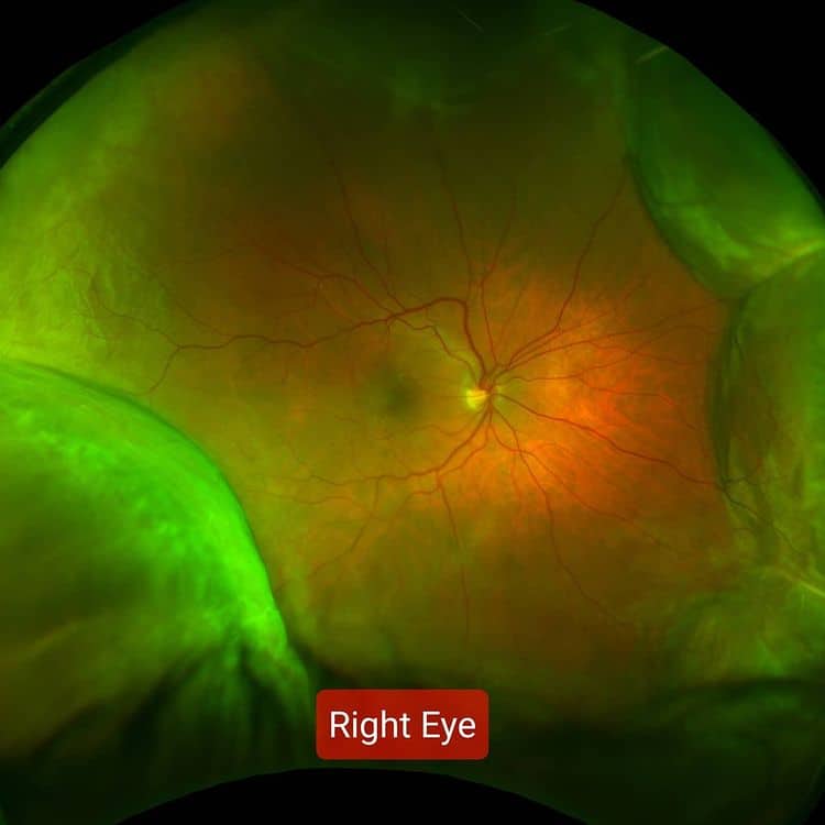
The walls are caving in..This 58yo male woke up with painless visual disturbances in both eyes. There was no preceding events or illness to note and his ocular and medical history was unremarkable. On examination, visual acuity was BE 6/12, his anterior segment was quiet and IOPs were normal. Posterior examination revealed circumferential choroidal detachments […]
Perifoveal Exudative Vascular Anomalous Complex (PEVAC)
It’s not always diabetes.. This 47yo male patient was referred for a second opinion for CME/CMO in his LE. He had a 6 month history of mild blurring and distortion. Ocular and medical history was unremarkable. He denies any cardiovascular risk factors including diabetes, hypertension, hyperlipidaemia and smokingOn examination his vision was RE 6/5, LE […]
Four Flange Fixation with Canabrava Technique
Following on from the previous video of the Four Flange Fixation with Canabrava technique, we demonstrate a couple of different ways to externalise the sutures. In this case, we are already using 27g ports for a preceding 27g vitrectomy. The different techniques are: 1) Preloading the suture within 27g needle as described by @pedror.henriques2) Docking […]
Canabrava Four-Flange IOL Fixation
Here’s an edited video of 4-Flange Fixation using the Canabrava technique initially described by @dr_canabrava . We like this technique as it combines the best attributes of the Yamane flange formation with 4-Point fixation but without the use of Goretex suture. This eliminates the need to rotate knots and any concerns about erosion or secondary granulomas from […]
Psoriatic Uveitis
Not Just a RashThis is a 60yo lady presenting with blurred vision and floaters RE for 1 week. There’s no significant ocular history and she’s medically well besides Psoriasis. Anterior examination revealed vision of RE 6/7.5, LE 6/6 with minor AC cells. Posteriorly she had bilateral vitritis RE>>LE. However, the most striking features were […]
CZ70BD Rescue with Cowhitch Knot
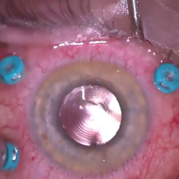
This is from a series of dislocated CZ70BD intraocular lenses (IOLs) that were rescued using 8-0 Goretex and employing a Cowhitch knot with 4-point fixation. The CZ70BD IOLs were traditionally sutured to sclera using 10-0 Prolene which has a limited lifespan of approximately 10-15 years before breaking down. Therefore many of these patients may require […]
Sequential Paracentral and Central Scotomas
Double Trouble!!This 60yo lady had sequential paracentral and central scotomas following trivial trauma and valsalva events. She initially had large multilayered bleeds in both eyes (images 1 &2) and underwent vitrectomy in the left eye for central sub-ILM bleed overlying her macula seen on OCT imaging (images 3&4), fortunately there was no subfoveal blood in […]
Progressive Visual Loss RE
Here’s an interesting case of a 14yo male referred in with progressive visual loss RE for 3 months and new onset central visual loss LE 1 week. Images 1 and 2 show colour photos of the pathology at 2 different time points of the same condition – with the RE illustrating chronic changes whereas […]
Paracentral Scotoma
Spot diagnosis! A 25yo female with a came in with a 2 day history of paracentral scotoma in the right eye on background of a recent viral illness (not COVID-19). Examination was unremarkable with VA RE 6/7.5 (20/25), LE 6/6 (20/20) and no evidence of inflammation anteriorly or posteriorly. She draws out a perfect […]
Combined Scleral Fixation of IOL and ERM Peel
Here’s a case of one of our surgeons performing a combined scleral fixation of a subluxated IOL with an ERM peel. This patient presented with a symptomatic dense epiretinal membrane (ERM) and a subluxated IOL with the optic edge close the visual axis. Given the high risk of IOL dislocation, the patient was consented for […]
Intraocular Pressure (IOP)
Case of the Week – Jump into the fog Here’s a 29yo female South American traveller referred in for left eye discomfort for 3 days and an initially raised intraocular pressure (IOP) of 48mmHg that has been treated by the referrer. On examination of the left eye, she had cells in the anterior chamber […]
Giant Retinal Tear (GRT)
Here’s a recent video of one of our surgeons repairing a pseudophakic macula-on Giant Retinal Tear (GRT) with pars plana vitrectomy. The steps are highlighted and despite having a diffractive multi-focal IOL, peripheral view remained excellent throughout the case. The use of Perfluorocarbon heavy liquid as a surgical device has greatly improved surgical […]
Angioid Streaks
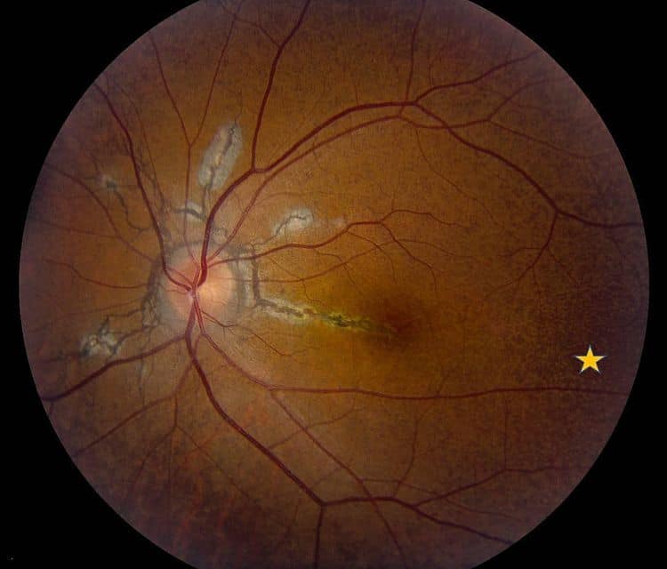
Our first “Case of the Week”!!These are colour photos and fluoroscein angiograms of a 30yo female who presents with progressive acuity loss OS. On examination she has Angioid Streaks (AS) radiating from the disc and is now involving her macula OS, hence the deteriorating vision.She also has Peau d’Orange appearance of the fundus (yellow stars) […]

