1")
 2")
 3")
 4")
 5")
 6")
 7")
Case of the Week – Jump into the fog
Here’s a 29yo female South American traveller referred in for left eye discomfort for 3 days and an initially raised intraocular pressure (IOP) of 48mmHg that has been treated by the referrer.
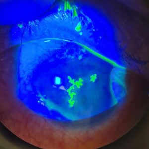
On examination of the left eye, she had cells in the anterior chamber and anterior vitreous. Fundoscopy of the left eye showed a new pale area of chorioretinitis adjacent to a major vessel and previous toxoplasmosis scar in the superotemporal quadrant. Right eye examination was unremarkable
Multimodal imaging demonstrates the hazy vitreous in Image 2 as well as the “headlight in fog” appearance of the lesion.
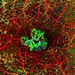
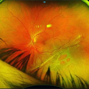
The angiogram (images 3 &4) demonstrates the choroidal non-perfusion of the lesion (image 3) as well as the choroidal blockage due to pigmentation of the existing scar. It also shows disc leakage and lowgrade vasculitis (image 4).
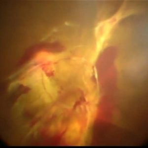
SD-OCT B images (5&6) show the thickened, raised and hyperreflective choroid, disorganised retina with overlying vitritis. The scans beautifully show the chorioretinitis and vitritis interface.
This is patient has Toxoplasmosis Chorioretinitis, confirmed on PCR through an AC tap.
On further questioning, the patient has a long history of pet cats.
She responded well to combined treatment of Bactrim and steroids (systemic and topical).
retina #ophthalmology #vitreoretinalsurgery #eyedisease #retinalsurgery #eyesurgery #ophthalmologist #eyedoctor #optometry #optometrist #optometrystudent #vitrectomy #macula #laser #toxoplasmosis #medicalretina #macula #laser #optomap #toxoplasmosis