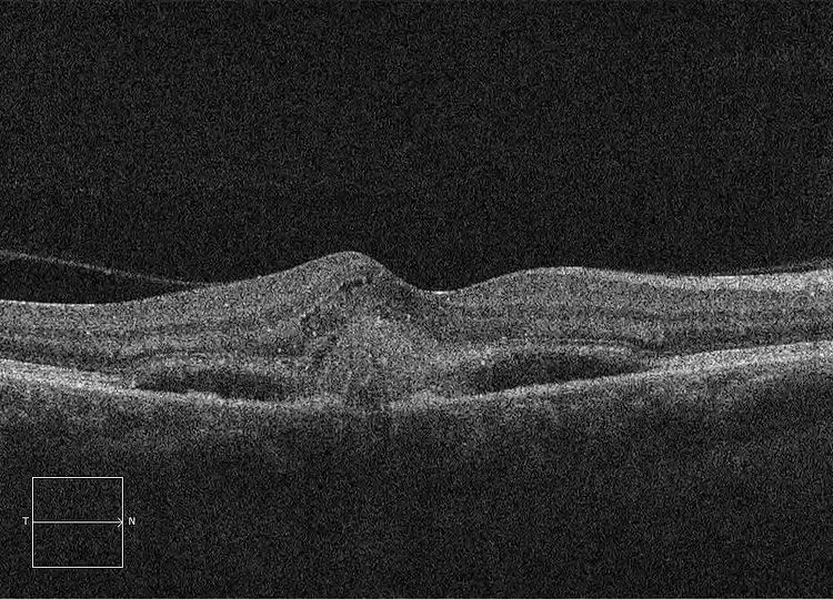
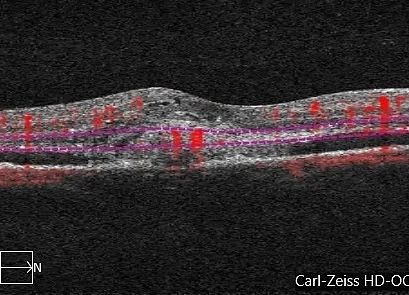
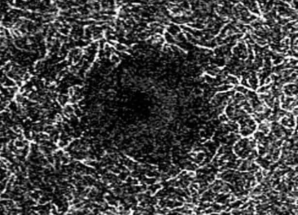
Here’s a series of OCTs and OCTAs of a neovascular (wet) Age-Related Macular Degeneration (nARMD) patient who underwent treatment with intravitreal anti-VEGF injections of Aflibercept.
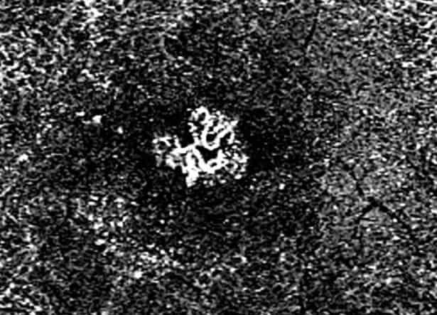
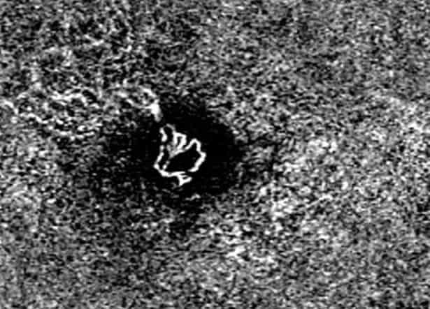
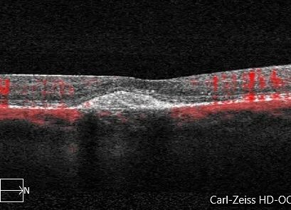
The initial (2nd image) OCT cross-sectional (B-scan) demonstrates subretinal and intraretinal fluid (SRF & IRF) with subfoveal haemorrhage. The third image is the OCT B-scan with flow overlay showing vascular flow in the subfoveal space. The horizontal lines demonstrate the flow is in the Outer Retina slab. The 4th image is the enface of the deep slab clearly showing the type 2 choroidal neovascular membrane (CNVM). The fourth image is the colour-coded enface angiogram.
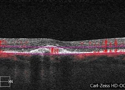
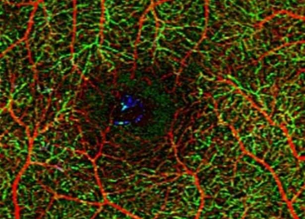
The 5th image shows improvement of the CNV on the ORCC and almost complete resorption of the subretinal and intraretinal oedema. This was following their first intravitreal injection. The 6th image shows the corresponding en-face OCTA.
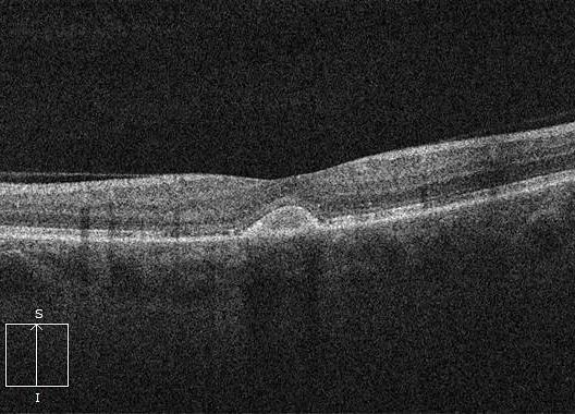
The 7th image demonstrates resolution of the CNV following 3 injections, with all that remains being a fibrous scar. This patient’s vision improved from 6/36 to 6/9 and has remained stable with ongoing treatment.
This series demonstrates both the effectiveness of treatment and utility of OCT Angiography in diagnosing disease and monitoring treatment progression.
#ophthalmology #ophthalmologist #retina #retinopathy #vitreoretinalsurgery #retinasurgery #vitrectomy #eyedisease #eyesurgery #eyedoctor #optometry #optometrist #optometrystudent #macula #maculopathy #laser #medicalretina #medret #medretina #octa #oct #cmo #cme #maculardegeneration #goldcoast #goldcoasteyes #outlookeye #goldcoasteyesurgeon #goldcoastoptom #multimodalimaging


