Spot diagnosis!
A 25yo female with a came in with a 2 day history of paracentral scotoma in the right eye on background of a recent viral illness (not COVID-19). Examination was unremarkable with VA RE 6/7.5 (20/25), LE 6/6 (20/20) and no evidence of inflammation anteriorly or posteriorly.
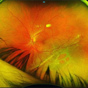
She draws out a perfect wedge on the Amsler (image 3) and on Near Infrared (NIR) a corresponding hyporeflective wedge/petaloid pointing towards the fovea can be visualised. This is a hallmark feature of Acute Macular Neuroretinopathy (AMN) .
The OCT-B scan shows the classic appearance of AMN with paracentral outer retinal hyperreflectivity especially OPL, thinning of ONL and disruption of the EZ&ELM.
These patients usually self resolve with no treatment necessary. Our patient’s symptoms resolved within 4 weeks with no residual effects.
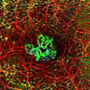
The final 2 images are from another recent case of bilateral AMN with multiple petals noted in each eye.
#retina #ophthalmology #vitreoretinalsurgery #eyedisease #retinasurgery #eyesurgery #ophthalmologist #eyedoctor #optometry #optometrist #optometrystudent #macula#amn #acutemacularneuroretinopathy #vitrectomy #retinaldetachment #macula #laser #goldcoast #retinalsurgery #eyedisease #eyesurgery #eyehealth #eyedoctor #medstudent #medicalstudent #goldcoast #goldcoasteyes #outlookeye #goldcoasteyesurgeon #goldcoastoptom