Here’s an interesting case of a 14yo male referred in with progressive visual loss RE for 3 months and new onset central visual loss LE 1 week.
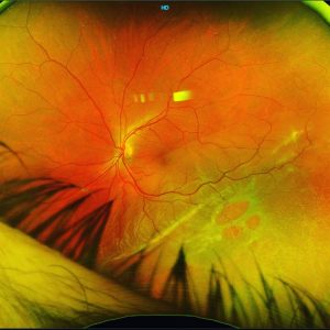
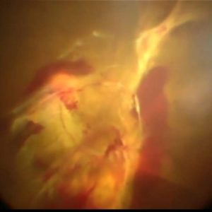
Images 1 and 2 show colour photos of the pathology at 2 different time points of the same condition – with the RE illustrating chronic changes whereas the LE demonstrating acute retinal whitening. The linear streaks on the colour images 1 and 2 are almost pathognomonic of this condition and the curvilinear hyperreflective bands along Henle’s layer on OCT of the LE (image 5) provide further evidence. Images 3 & 4 demonstrate the hyperautofluorescent streaks with the acute findings, mostly on the LE.
Fluorescein angiography (image 6) as well as OCT angiography (image 7) show a secondary choroidal neovascular membrane in the RE.
On further questioning, the patient admitted to repeated self exposure to a high-powered Handheld Laser purchased online.
With only a single aflibercept injection followed by close observation for 6mths this patient went from 6/120 to 6/9 in the RE (?!!).
This is a fairly classic example of Handheld Laser Induced Maculopathy (HLIM), a condition on the rise internationally due to their online availability.
We had 2 HLIM cases referred for second opinions in 1 month. Anyone else notice an increase in these presentations??
What’s your preferred management? Steroids? Observation?
#retina #ophthalmology #vitreoretinalsurgery #eyedisease #retinasurgery #eyesurgery #ophthalmologist #eyedoctor #optometry #optometrist #optometrystudent #vitrectomy #retinaldetachment #macula #laser #goldcoast #goldcoasteyes #outlookeye #goldcoastoptom #laser #hlim #lasermaculopathy #lasers #medicalretina #angiography #octangiography #octa #oct #retinopathy #maculopathy #cme