Here’s a series of photos from separate patients demonstrating the 2 types of Retinal Capillary Haemangiomas – Peripheral and Juxtapapillary.
Retinal Capillary Haemangiomas are vascular hamartomas that can not only cause slowly progressive visual problems but may also be associated with Von-Hippel Lindau (VHL) disease.
RCHs can be isolated, multiple or bilateral. Peripheral RCHs appear as small red cherry-like mass between large, dilated, tortuous arterioles and venules extending from the optic disc to the RCH (image 1&2).
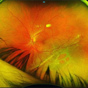
Juxtapapillary RCHs are not associated with arteriovenous shunts. They are also usually red/pink intraretinal lesions and may be associated with oedema, (simulating optic nerve oedema) and juxtapapillary choroidal neovascularization and bleeds (image 3&4). Although they are benign, slowly growing tumours, juxtapapillary RCHs can cause significant visual deterioration secondary to progressive intraretinal and subretinal exudation, macular edema and exudative retinal detachment if left untreated.
RCHs have 3 possible growth patterns. Endophytic lesions originate in the inner retinal layers and grow into the vitreous cavity, therefore are easily identifiable. Sessile tumours originate within the middle retinal layers and appear as grey-orange flat lesions. Exophytic tumors grow in the outer layers of the retina and present clinically as orange, nodular well-defined lesions.
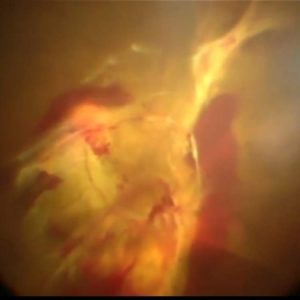
Fluorescein Angiography is useful to show progressive hyperfluorescent leakage of juxtapapillary intraretinal lesions and to differentiate it from optic nerve oedema and choroidal neovascularization (images 5-7). It is also useful to detect arteriovenous shunting in peripheral endophytic tumors (image 2) , and to detect very small lesions.
#ophthalmology #ophthalmologist #retina #retinopathy #vitreoretinalsurgery #retinasurgery #vitrectomy #eyedisease #eyesurgery #eyedoctor #optometry #optometrist #optometrystudent #macula #maculopathy #laser #medicalretina #medret #medretina #angiography #octa #oct #cmo #cme #maculardegeneration #goldcoast #goldcoasteyes #outlookeye #goldcoasteyesurgeon #goldcoastoptom