This young 27yo female presents with sudden loss of vision in the right eye.
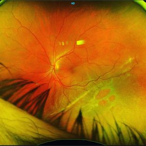
Vision dropped to 6/12 (20/40). Anterior examination was unremarkable. Posterior examination revealed large areas of retinal whitening emanating from the optic disc of the right eye involving the macula.
This patient has Serpiginous Choroidopathy, which is part of the Placoid spectrum marked by geographic choroiditis or peripapillary choroidopathy. Patients present wth
characteristic serpent-like unifocal, grayish lesions located peripapillary or starting at macula (10% of cases).
It’s a rare inflammatory choroiditis of unknown cause with secondary affects on outer retina and RPE .
It is usually bilateral/sequential, asymmetric, recurrent/chronic. Typical course is marked by progression , regression and recurrences which may occur months to years apart.
Fluorescein angiography findings include:
• Early hypofluorescence due to choriocapillaris atrophy/hypoperfusion
• Progressive hyperfluorescence at lesion margins
• Late staining/leak of active border and underlying sclera and fibrosis
Fundus Autofluorescent findings include:
• Hyperfluorescent active lesions usually appear 2-5 days after appearance of lesions delineating are of RPE damage
• Hypofluorescent regressed/atrophic lesions
OCT findings include:
• Outer retinal hyperreflectivity and choroidal thickening in active lesions.
• Retinal and RPE atrophy in regressed lesions.
Treatment involves high dose steroids as well as long term immunosuppresion co-managed with the rheumatologist.
#retina #ophthalmology #vitreoretinalsurgery #eyedisease #eyespecialist #retinasurgery #eyesurgery #ophthalmologist #eyedoctor #ophthalmologyresident #optometry #optometrist #optometrystudent #vitrectomy #macula #maculopathy #retinopathy #laser #uveitis #serpiginous #optomap #medicalretina #medret #goldcoastoptom #goldcoasteyes #outlookeye #goldcoasteyesurgeon