1")
 2")
 3")
 4")
 5")
 6")
Here’s a case of X-Linked Retinoschisis (XLRS)
This is an inherited disorder affecting males, generally diagnosed in childhood due to an inherited mutation in RS1 gene (which encodes for a protein involved in intercellular adhesion and organisation – Retinoschisin).
It leads to bilateral reduced central vision which may result in nystagmus or strabismus. Vision generally stabilises in early adulthood between 20/60 and 20/120 though it may further deteriorate after the 6th decade. Patients may also lose vision from significant complications including vitreous bleeds (<30%) due to rupture of vessels crossing the schisis cavities or retinal detachments (5-20%).
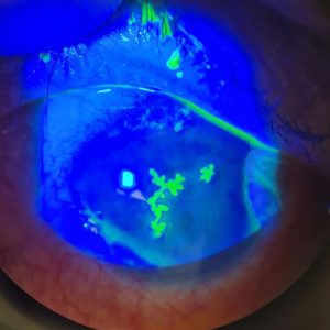
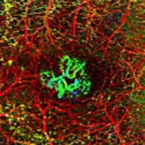
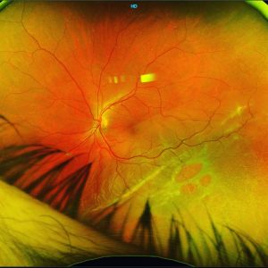
Patients present with characteristic foveal schisis which appears as central radial spokewheel pattern, often better visualised on red-free. Peripheral retinoschisis is present in 50% of patients
OCT may demonstrate splitting of several layers of the retina, howeve the inner layers split more commonly. OCT may also exhibit large central cysts.
Fluorescein angiography can be used to differentiate between cystic macular oedema as XLRS has an abscence of petaloid leakage.
To date, there are no available treatments to stop XLRS progression. Surgery is required for any non-clearing or amblyogenic vitreous bleeds or retinal detachments.
#medicalretina #medret #medretina #angiography #octa #oct #multimodalimaging #retinopathy #maculopathy #cmo #cme #maculardegeneration #ophthalmology #ophthalmologist #retina #vitreoretinalsurgery #retinasurgery #vitrectomy #eyedisease #eyesurgery #eyedoctor #optometry #optometrist #optometrystudent #macula #goldcoast #goldcoasteyes #outlookeye #goldcoasteyesurgeon #goldcoastoptom